...

6 Exercise Phenylketonuria

You need to read chapters “Protein digestion and Amino acid absorption” and “Amino acid metabolism” before attempting this tutorial. In the following tutorial four different mouse strains are used. 

All images are from : Belanger AM, Przybylska M, Gefteas E, Furgerson M, Geller S, Kloss A, Cheng
SH, Zhu Y, Yew NS. Inhibiting neutral amino acid transport for the treatment of phenylketonuria. JCI Insight. 2018 Jul 26;3(14). pii: 121762. doi: 10.1172/jci.insight.121762

The table below shows the mouse strains used in this study.

Strain
Physiological phenotype
wt
Normal mouse strain (control)
SLC6A19
Mouse strain lacking SLC6A19 a transporter
for neutral amino acids that is mutated in Hartnup disorder.
Pahenu2
A mouse strain with a mutation
in phenylalanine hydroxylase (PAH)
Pahenu2 x SLC6A19 HET
A cross between SLC6A19 and PAHenu2,
PAH is non-functional, one allele of SLC6A19 is functional.
Pahenu2 x SLC6A19 KO
A homozygous cross between SLC6A19 and
PAHenu2, both proteins are non-functional.

Question 1: What is Pheylketonuria?

Phenylketonuria (PKU) is a rare disorder of amino acid metabolism. It is caused by mutations in the enzyme phenylalanine hydroxylase. This enzyme converts phenylalanine to tyrosine. It is the first step of phenylalanine breakdown. Tyrosine metabolism is unaffected.

Question 2: What is the hallmark of phenylketonuria? Is it replicated in mice with mutations in phenylalanine hydroxylase (Pahenu2, Fig. 1)?

Phenylalanine levels in urine, plasma and brain of different mouse strains.

Plasma phenylalanine levels are highly elevated in PKU.

Question 3: Why are phenylalanine levels in urine elevated when SLC6A19 is mutated? What happens when there is also a mutation in phenylalanine hydroxylase?

After filtration in the glomerulus, amino acids including phenylalanine are reabsorbed. Transporters are required to mediate the re-uptake of amino acids, such as SLC6A19, which transports neutral amino acids.

Question 4: Why are urine phenylalanine levels not elevated in the PAHenu2 mice?

Although high plasma Phenylalanine levels translate to very high levels of Phenylalanine in the glomerular filtrate the capacity of transport must be high enough to remove all Phenylalanine.

Question 5: What happens to plasma amino acid levels in the other strains?

Wildtype shows physiological levels. PAHenu2 has highly elevated levels because phenylalanine cannot be metabolised and accumulates. The PAHenu2 x SLC6A19KO shows levels that are closer to wildtype again, because Phenylalanine is released through the urine. The PAHenu2 x SLC6A19 HET does not improve the condition, because Phenylalanine does not spill over into the urine.

Phenylketonuria (PKU) causes mental retardation if untreated. It is assumed that neurotransmitter levels and neurotransmitter metabolism in the brain are affected in PKU. This can be demonstrated in a mouse model of PKU (Fig. 2). Dopamine and Norepinephrine are derived from Phenylalanine or Tyrosine, while Serotonin is derived from Tryptophan. HIAA (Hydroxyindoleacetic acid) is a breakdown product of Tryptophan.

Fig. 1 Brain content of neurotransmitters in different mouse strains. 5-HIAA 5-hydroxyindolacetic acid, a metabolite of serotonin.

Question 6: Why are neurotransmitter levels lower in the brain of PKU mice?

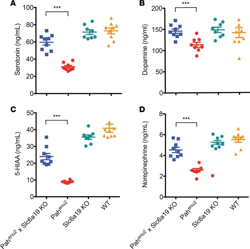
There are several possibilities. Phenylalanine could compete with tyrosine for the next metabolic steps in the generation of dopamine and noradrenaline, but this would not affect synthesis of serotonin, which is derived from tryptophan. More likely phenylalanine competes with tryptophan and tyrosine transport into the brain. Although tyrosine is synthesized from Phenylalanine by PAH, tyrosine levels are normal when nutrition is adequate. 

Damage to neurons in the brain can be detected because of astrocytes (another cell type in the brain that supports neurons) becoming reactive and expressing GFAP (Fig. 3).

Fig. 3 Decreased number of reactive astrocytes in the ventral pallidum region of Pahenu2 mice lacking Slc6a19 compared with Pahenu2 mice. Brains from the different genotypes of mice were sectioned and stained with an anti-GFAP antibody, and the number of immunoreactive astrocytes was counted. Arrows indicate representative dark brown–stained, star-shaped astrocytes. Scale bar: 50 μm. n = 5 animals/group. One-way ANOVA was performed, follow by Tukey’s multiple comparisons test. **P = 0.003. Mean ± SEM is shown.

The occurrence of reactive astrocytes indicates brain injury. Interpret the results.

Less brain injury when phenylalanine levels are normalised.

Reduced brain development can be measured by investigating the complexity of the neuronal network. This can be done by counting branches or dendritic spines where neurons connect to each other.

Fig. 4 Dendritic spine density and arborization complexity in different mouse strains. (A) Dendritic spines were visualized using Golgi-Cox staining. Shown are representative images from the CA1 and dentate gyrus regions of the hippocampus and layers 2/3 of the medial prefrontal cortex. A minimum of 750 spines from 5 neurons per mouse and at least 3 mice per genotype were analyzed. Representative neurons from Pahenu2, wild-type, and Pahenu2 × Slc6a19-KO mice from layers 2/3 of the cortex were visualized using Golgi-Cox staining and electronically traced using Neurolucida Suite and Cell Sens software. Original magnification, ×1,000. (B) Pahenu2 × Slc6a19-KO neuron. (C) Pahenu2 neuron. (D) Wild-type neuron. (E) Quantification of branch number. n = 16–20 neurons analyzed per genotype. (F) Quantification of branch complexity. Branch complexity was quantified using a priming scheme, with branches directly connected to the neuronal cell body identified as 1′, secondary branch points identified as 2′, etc. One-way ANOVA was performed, follow by Tukey’s multiple comparisons test. **P < 0.005, ***P < 0.001. Mean ± SEM is shown.

Task: Comment on the differences of brain development in the mouse strains.

Cognition and brain function relies on a highly developed neural network. The number of nerve inputs can be derived from the number of dendritic spines. Hi Phenylalanine levels delay brain development.

Question: What is the significance of this study?

The study demonstrates the feasibility of a pharmacological treatment of PKU through blocking the renal and intestinal amino acid transporter SLC6A19. Drugs are now developed to achieve this aim.

https://insight.jci.org/articles/view/182876

Seraphinite AcceleratorOptimized by Seraphinite Accelerator
Turns on site high speed to be attractive for people and search engines.